Please enter an email address
This website will not save the email address. It will only be used to send the requested document.

This website will not save the email address. It will only be used to send the requested document.
You are about to go to a website sponsored by Amgen. Would you like to continue?
You will be going to an external website operated by an independent third party. Your activities at the external website will be managed by their policies and practices. By clicking “Continue,” you will go to the external website.
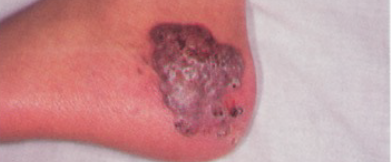
Review the radiographic images below to better understand how infections and granulomas may present in patients with CGD.
Autoimmune disorders are common in CGD. Some autoimmune diseases reported in individuals with CGD include juvenile idiopathic arthritis, autoimmune pulmonary disease, and myasthenia gravis.1


Osteomyelitis in a patient aged 4 years with CGD who presented with a limp.3


8-year-old patient with CGD presenting with pulmonary infection, fever, and respiratory distress.3


Early diagnosis is beneficial for patients with CGD because they are susceptible to serious infections.1,4* If CGD is suspected, a dihydrorhodamine (DHR) test can confirm a diagnosis.

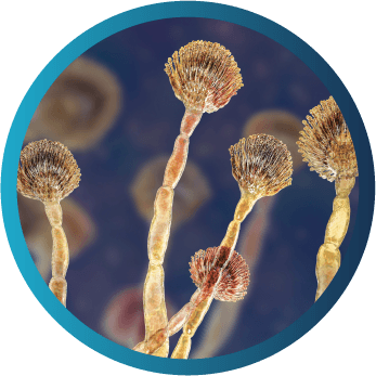
Many severe infections in patients with CGD in North America are caused by a select group of organisms, both bacterial and fungal.1,5-10 Most of these pathogens, including Aspergillus, Nocardia, Serratia, Burkholderia, Klebsiella, and Staphylococcus aureus, are catalase-positive.5,6 In patients with CGD, these catalase-positive pathogens are noteworthy because they can neutralize some of the hydrogen peroxide produced by neutrophils other than those in nicotinamide adenine dinucleotide phosphate (NADPH) oxidase.6
Common presentation: pneumonia, lymphadenitis, osteomyelitis, brain abscess

Common presentation: sepsis, soft tissue infection, liver abscess
*Serious infection is defined as a clinical event requiring hospitalization and/or intravenous antibiotics.
†This is not a complete list of pathogens. Infections may also be caused by other species of bacteria and fungi not listed here.
‡Candida is most commonly reported in the EU.
Staphylococcus aureus, Burkholderia cepacia complex, Serratia marcescens, and Nocardia species are the primary causes of most infections in individuals with CGD.1,9

Common presentation: pneumonia, osteomyelitis, brain abscess

Common presentation: osteomyelitis, soft tissue infections; less common presentation: pneumonia, sepsis

Common presentation: pneumonia, sepsis

Common presentation: pneumonia, skin infections, lymphadenitis

Common presentation: soft tissue infections, lymphadenitis, liver abscess, perirectal abscess, osteomyelitis, pneumonia, sepsis
*Serious infection is defined as a clinical event requiring hospitalization and/or intravenous antibiotics.
†This is not a complete list of pathogens. Infections may also be caused by other species of bacteria and fungi not listed here.
‡Candida is most commonly reported in the EU.
1. Leiding JW, Holland SM. Chronic granulomatous disease. In: Pagon RA, Adam MP, Ardinger HH, et al, eds. GeneReviews®. Washington, Seattle: University of Washington, Seattle; 1993-2022. 2. Dohil M, Prendiville JS, Crawford RI, Speert DP. Cutaneous manifestations of chronic granulomatous disease. A report of four cases and review of the literature. J Am Acad Dermatol. 1997;36(6 pt 1):899-907. 3. Khanna G, Kao SC, Kirby P, Sato Y. Imaging of chronic granulomatous disease in children. Radiographics. 2005;25(5):1183-1195. 4. Holland SM. Chronic granulomatous disease. Clin Rev Allergy Immunol. 2010;38(1):3-10. 5. Bonilla FA, Khan DA, Ballas ZK, et al. Practice parameter for the diagnosis and management of primary immunodeficiency. J Allergy Clin Immunol. 2015;136(5):1186-1205.e1-e78. 6. Bortoletto P, Lyman K, Camacho A, et al. Chronic granulomatous disease: a large, single-center US experience. Pediatr Infect Dis J. 2015;34(10):1110-1114. 7. Leiding JW, Malech HL, Holland SM. Clinical Focus on Primary Immunodeficiencies: Chronic Granulomatous Disease. Immune Deficiency Foundation; 2013. 8. Marciano BE, Zerbe CS, Falcone EL, et al. X-linked carriers of chronic granulomatous disease: Illness, lyonization, and stability. J Allergy Clin Immunol. 2018;141(1):365-371. 9. Song E, Jaishankar GB, Saleh H, Jithpratuck W, Sahni R, Krishnaswamy G. Chronic granulomatous disease: a review of the infectious and inflammatory complications. Clin Mol Allergy. 2011;9(1):10-24. 10. van den Berg JM, van Koppen E, Ahlin A, et al. Chronic granulomatous disease: the European experience. PLoS One. 2009;4(4):e5234.